Pelvic Anatomy Female Ligaments : Surgical Anatomy Of The Female Pelvis By Laparoscopy / These ligaments also play a crucial role in pelvic organ prolapse with anterior vaginal wall descent (5).
Pelvic Anatomy Female Ligaments : Surgical Anatomy Of The Female Pelvis By Laparoscopy / These ligaments also play a crucial role in pelvic organ prolapse with anterior vaginal wall descent (5).. Ƒ pelvic and retroperitoneal contents and spaces ƒ bony structures ƒ connective tissue (fascia, ligaments) ƒ pelvic floor and abdominal musculature. The pelvic floor muscles include; It allows flexion and extension of the coccyx. The sacrospinous ligament spans the sacrum to the ischial spine, and the sacrotuberous ligament spans the sacrum to the ischial tuberosity. The female bony pelvis is divided into:
This life size three part model represents an original cast of a bony female pelvis with ligaments. The axial (horizontal) image of the female pelvis shows the ovaries, uterus, ligament, uterine tubes, vaginal cavity, and other internal organs. Inguinal ligament, sacrotuberous ligament, sacrospinous ligament, anterior sacroiliac ligaments, iliolumbar ligament, anterior longitudinal ligament, interosseous sacroiliac ligament, posterior sacroiliac ligament and obturator. Ƒ vascular supply ƒ neurologic supply. This image shows the posterior back view of the female pelvic brim (the bones and ligaments that forms the pelvic region in the female) showing:
Female Pelvic Anatomy Flashcards Chegg Com from media.cheggcdn.com • anterolateral wall—hip bone and obturator internus muscles. • also known as pelvic cavity. This life size three part model represents an original cast of a bony female pelvis with ligaments. The pelvis is held together by three principal ligaments: The bony pelvis in normal standing posture transmits the body weight of head, trunk and the upper extremities to the lower extremities. The sacrococcygeal joint is an amphiarthrodial joint between the fifth sacral and first coccygeal segments. The pelvic floor muscles include; The sagittal (longitudinal) image of the female pelvis shows anatomical structures.
This life size three part model represents an original cast of a bony female pelvis with ligaments.
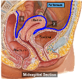
These ligaments also play a crucial role in pelvic organ prolapse with anterior vaginal wall descent (5). Functions of the pelvis the strong and rigid pelvis is adapted to serve a number of roles in the human body. The sagittal (longitudinal) image of the female pelvis shows anatomical structures. These ligaments are important stabilizers. Posts tagged female pelvic anatomy ligaments. Bones and ligaments of the female pelvis. The inlet to the pelvic canal is at the level of the sacral promontory and superior aspect of the pubic bones. The bony pelvis in normal standing posture transmits the body weight of head, trunk and the upper extremities to the lower extremities. • muscles and ligaments form a pelvic floor. In women, the ligaments of the joint soften during pregnancy, enabling the increase of pelvic diameter during childbirth. • lateral boundaries—fused ilium and ischium. • also known as pelvic cavity. The right half of the model shows the following pelvic ligaments:
Imaios and selected third parties, use cookies or similar technologies, in particular for audience measurement. Ligaments of the pelvis the posterior sacroiliac ligament supports the sacroiliac joint. The sacrococcygeal joint is an amphiarthrodial joint between the fifth sacral and first coccygeal segments. Functions of the pelvis the strong and rigid pelvis is adapted to serve a number of roles in the human body. The cardinal and uterosacral ligaments, which support the uterus and upper part of the vagina, are critical structures in the female pelvis (3, 4).
Usmle Step 1 Anatomy Female Ligaments And Local Structures Youtube from i.ytimg.com This anatomically detailed model is a great way to teach and learn the anatomy of the human female pelvis. The broad ligament ends where the infundibulopelvic ligament blends with the pelvic wall. Finally, a checklist is provided for structured reporting of the mri findings in the female pelvis. Pelvic anatomy sacrouterine ligament cardinal ligaments pelvic fascia sacrospinous ligament urethral support bladder support rectal support. Suspended in the mesovarium (attached to the posterior part of the broad ligament). The broad ligament can be further divided into three components that are linked to. The sacrococcygeal joint is an amphiarthrodial joint between the fifth sacral and first coccygeal segments. Cookies allow us to analyze and store information such as the characteristics of your device as well as certain personal data (e.g., ip addresses, navigation, usage or geolocation data, unique identifiers).
• posterolateral wall—piriformis and coccygeus muscles.
Ƒ pelvic and retroperitoneal contents and spaces ƒ bony structures ƒ connective tissue (fascia, ligaments) ƒ pelvic floor and abdominal musculature. Suspended in the mesovarium (attached to the posterior part of the broad ligament). This anatomically detailed model is a great way to teach and learn the anatomy of the human female pelvis. The sacrococcygeal joint is an amphiarthrodial joint between the fifth sacral and first coccygeal segments. Many pelvic landmarks, ligaments, and muscular structures within the pelvis are important to know to differentiate normal reproductive organs from muscular and vascular structures. Imaios and selected third parties, use cookies or similar technologies, in particular for audience measurement. Cookies allow us to analyze and store information such as the characteristics of your device as well as certain personal data (e.g., ip addresses, navigation, usage or geolocation data, unique identifiers). Also, the compartmental anatomy of the female pelvis is explained, including the extraperitoneal pelvic spaces. The obstetrical anatomy of a typical female pelvis is best considered as one unit. Ligaments of the pelvis the posterior sacroiliac ligament supports the sacroiliac joint. The female pelvis because of its characteristics, aids in child birth. The cardinal and uterosacral ligaments, which support the uterus and upper part of the vagina, are critical structures in the female pelvis (3, 4). The broad ligament supports the uterus, fallopian tubes, and ovaries.
The broad ligament ends where the infundibulopelvic ligament blends with the pelvic wall. The cardinal and uterosacral ligaments, which support the uterus and upper part of the vagina, are critical structures in the female pelvis (3, 4). Additional ligaments may be found in the female pelvis. The ligaments of the female reproductive tract can be divided into three categories: Bones and ligaments of the female pelvis.
Female Pelvic Anatomy Medical Exhibit Medivisuals from medivisuals1.com The broad ligament ends where the infundibulopelvic ligament blends with the pelvic wall. • lateral boundaries—fused ilium and ischium. This image is a longitudinal cross section in the female pelvic brim (the bones and ligaments of the female pelvic region) displaying a lateral view of that sectioned pelvis showing: Finally, a checklist is provided for structured reporting of the mri findings in the female pelvis. The inlet to the pelvic canal is at the level of the sacral promontory and superior aspect of the pubic bones. The sacrospinous and sacrotuberous ligaments contribute to the formation of the greater and lesser sciatic foramens. It can be described as one of the bodies diaphragms. The pelvis is held together by three principal ligaments:
The bony pelvis in normal standing posture transmits the body weight of head, trunk and the upper extremities to the lower extremities.
The axial (horizontal) image of the female pelvis shows the ovaries, uterus, ligament, uterine tubes, vaginal cavity, and other internal organs. There are two, one on each side of the uterus, each composed of a double layer of peritoneum. Finally, a checklist is provided for structured reporting of the mri findings in the female pelvis. Other ligaments attached to bony pelvis include the sacrococcygeal ligaments, pubic symphysis ligaments, and endopelvic fascia ligament. The outlet is formed by the pubic arch, ischial spines, sacrotuberous ligaments, and the coccyx. Also, the compartmental anatomy of the female pelvis is explained, including the extraperitoneal pelvic spaces. Evolvement •forms a bony ring through with body 20. In female it is adapted for child bearing. The broad ligament can be further divided into three components that are linked to. Inguinal ligament, sacrotuberous ligament, sacrospinous ligament, anterior sacroiliac ligaments, iliolumbar ligament, anterior longitudinal ligament, interosseous sacroiliac ligament, posterior sacroiliac ligament and obturator. • lateral boundaries—fused ilium and ischium. The obstetrical anatomy of a typical female pelvis is best considered as one unit. The function of the pelvic floor is to help assist with child birth, prevent incontinence and support organs within the pelvis.


0 Komentar